Osteoid Osteoma: Say Goodbye to Pain with Scarless CT-Guided RFA!
Osteoid Osteoma: Advanced Treatment with CT-Guided RFA
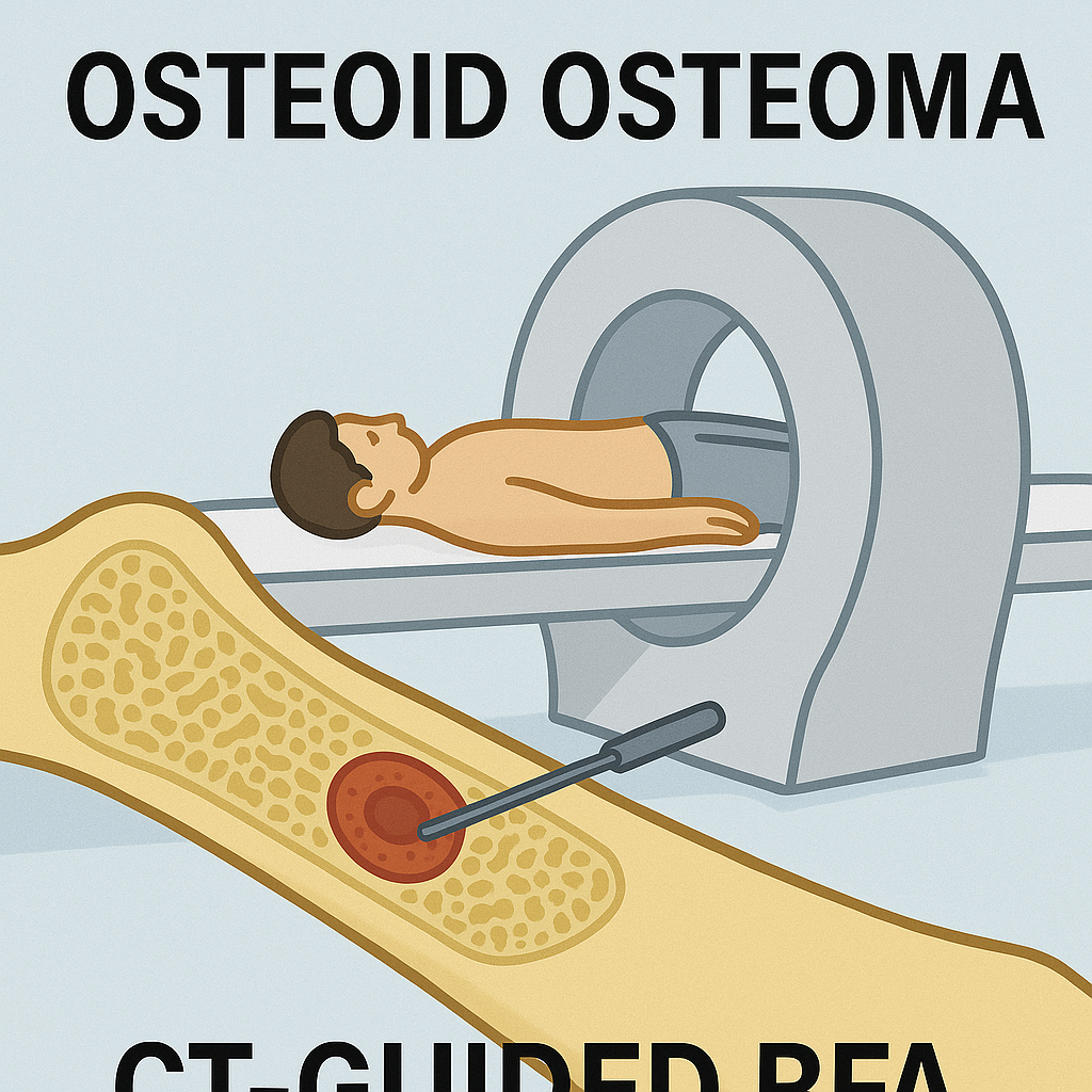
What is Osteoid Osteoma?
Osteoid osteoma is a benign but painful bone tumor that primarily affects young individuals, typically between the ages of 10 and 35. It commonly occurs in the long bones of the body, such as the femur and tibia, and causes persistent pain that worsens at night, often relieved by nonsteroidal anti-inflammatory drugs (NSAIDs).
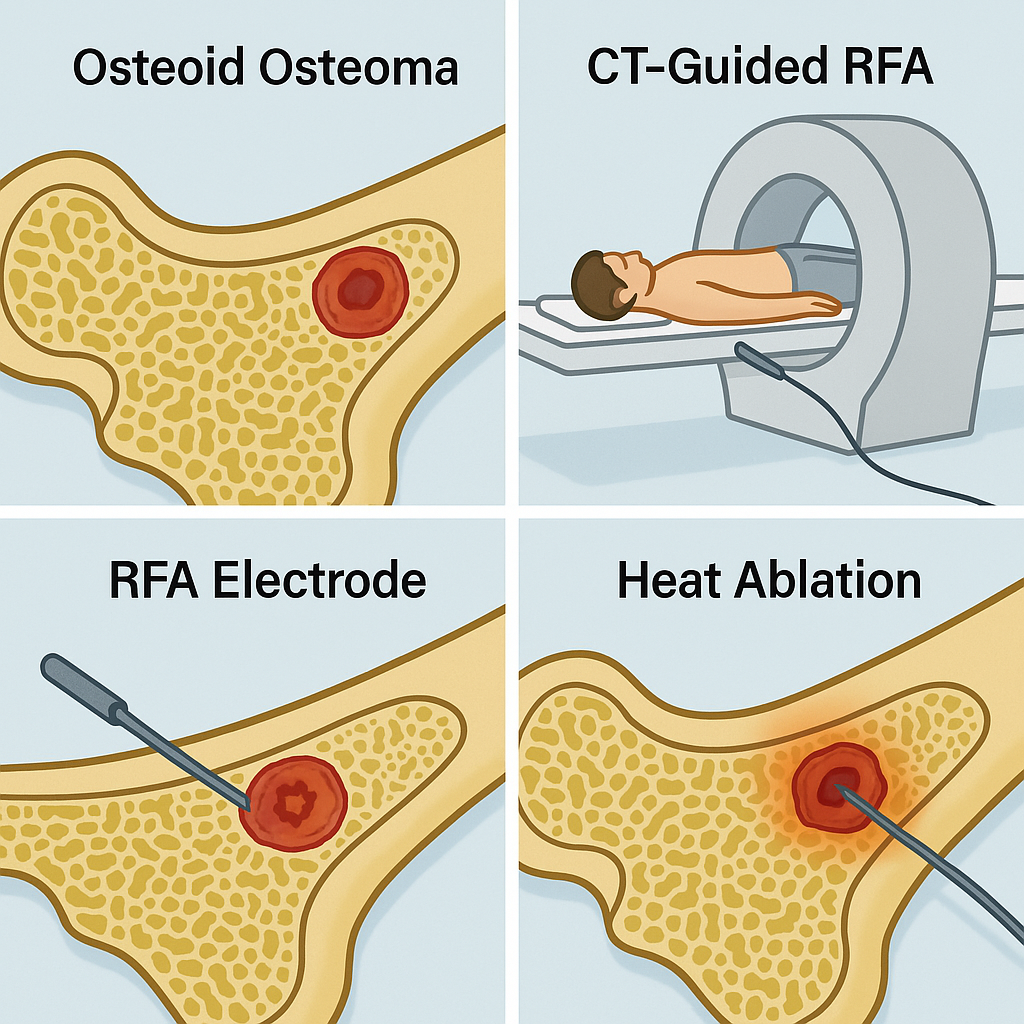
Treatment of Choice: CT-Guided Radiofrequency Ablation (RFA)
CT-guided Radiofrequency Ablation (RFA) is the gold standard and minimally invasive procedure for treating osteoid osteoma. Unlike traditional open surgery, RFA is a sutureless technique that offers excellent results with minimal complications.
Why Choose CT-Guided RFA?
- Minimally invasive: No major incisions or stitches required.
- High success rate: More than 95% effectiveness in completely destroying the tumor.
- Same-day discharge: Patients can return home within a few hours after the procedure.
- Quick recovery: Return to normal activities within a few days.
- Less pain and scarring: Compared to traditional surgery, RFA has minimal discomfort and no significant scars.
Dr. Lokesh Garg: Your Expert in Bone Tumor Management
If you or a loved one has been diagnosed with osteoid osteoma, you need an expert who specializes in orthopedic oncology. Dr. Lokesh Garg is a leading Orthopedic Surgeon, Joint Replacement Surgeon, and Ortho Onco Specialist, known for his expertise in treating bone tumors, including osteoid osteoma, using advanced techniques like CT-guided RFA. Dr. Lokesh Garg personally performs each of these procedures, ensuring precision, safety, and optimal outcomes for his patients.
Why Should You Avoid Getting This Procedure Done by an Interventional Radiologist?
While interventional radiologists may offer RFA, they lack the expertise in bone drilling and understanding of surrounding anatomical structures. This can lead to:
- Risk of improper needle placement: Without orthopaedic knowledge, incorrect placement may lead to ineffective treatment.
- Potential damage to surrounding structures: Heat from the RFA probe can injure nearby nerves, vessels, or muscles if not handled correctly.
- Inability to manage complications: If a complication arises, interventional radiologists may not be equipped to perform surgical interventions or manage post-procedure complications.
By choosing Dr. Lokesh Garg, you ensure that your procedure is performed by an orthopaedic expert who understands bone structures, uses precise drilling techniques, and can handle any unforeseen complications with ease.
Success Stories: Previous Cases Treated
We have successfully treated numerous patients with osteoid osteoma using CT-guided RFA. Here is a sample case template for previous treatments:
Case Template:
- Patient Age & Gender: 18 Month kid
- Symptoms: Night pain in left thigh for 3 months, relieved by NSAIDs
- Diagnosis: Confirmed osteoid osteoma through imaging and clinical assessment.
- Treatment: CT-guided Radiofrequency Ablation performed under general anaesthesia.
- Outcome: Immediate pain relief, no complications, and same-day discharge.
- Follow-up: Complete recovery with no recurrence after 6 months.
If you are experiencing persistent bone pain and suspect osteoid osteoma, consult Dr. Lokesh Garg today for an expert opinion and world-class treatment with CT-guided RFA.
📞 Contact Us Today for an appointment and take the first step toward pain-free living!